


De lactibus sive lacteis venis quarto vasorum mesaraicorum genere novo invento... dissertatio.
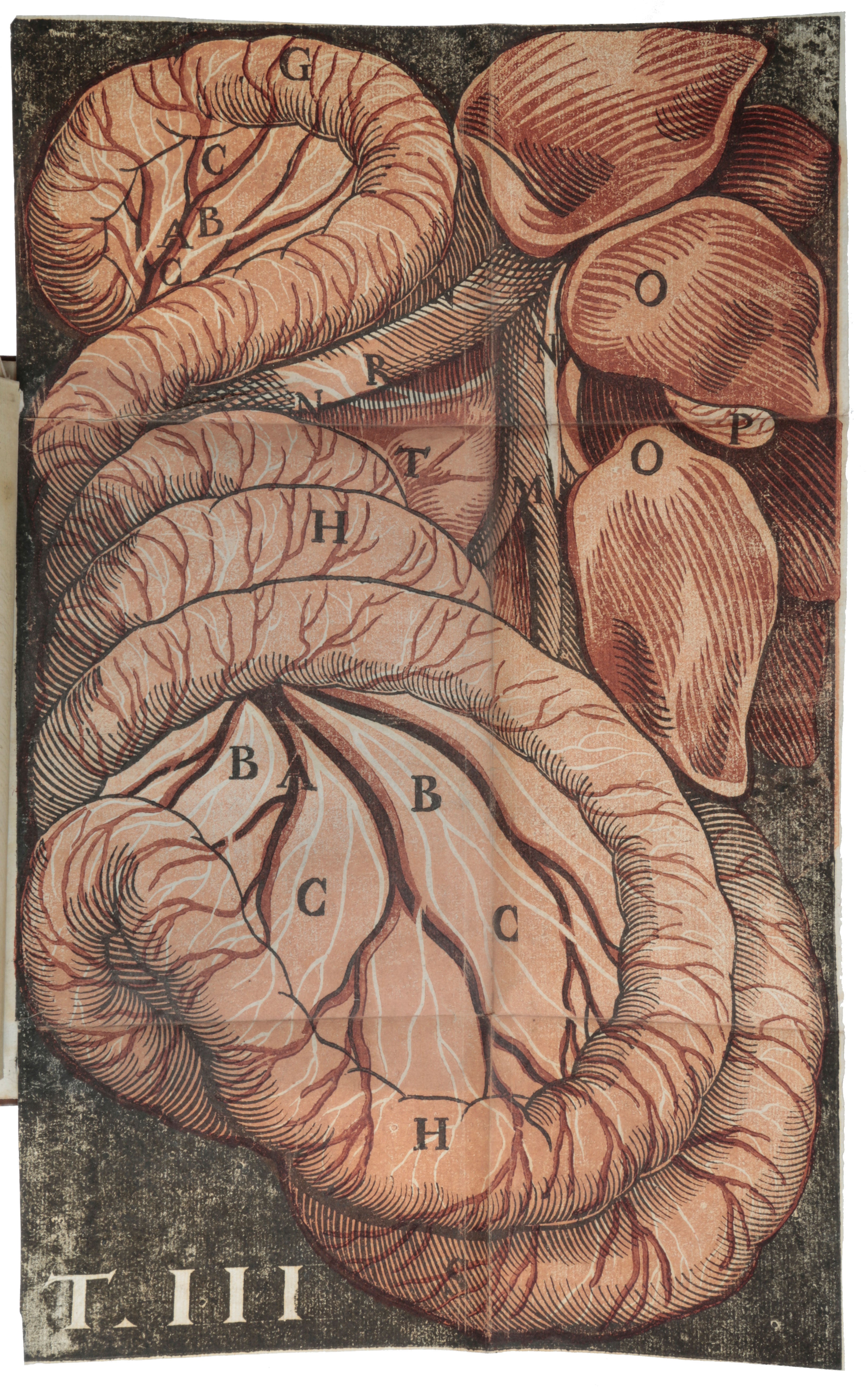
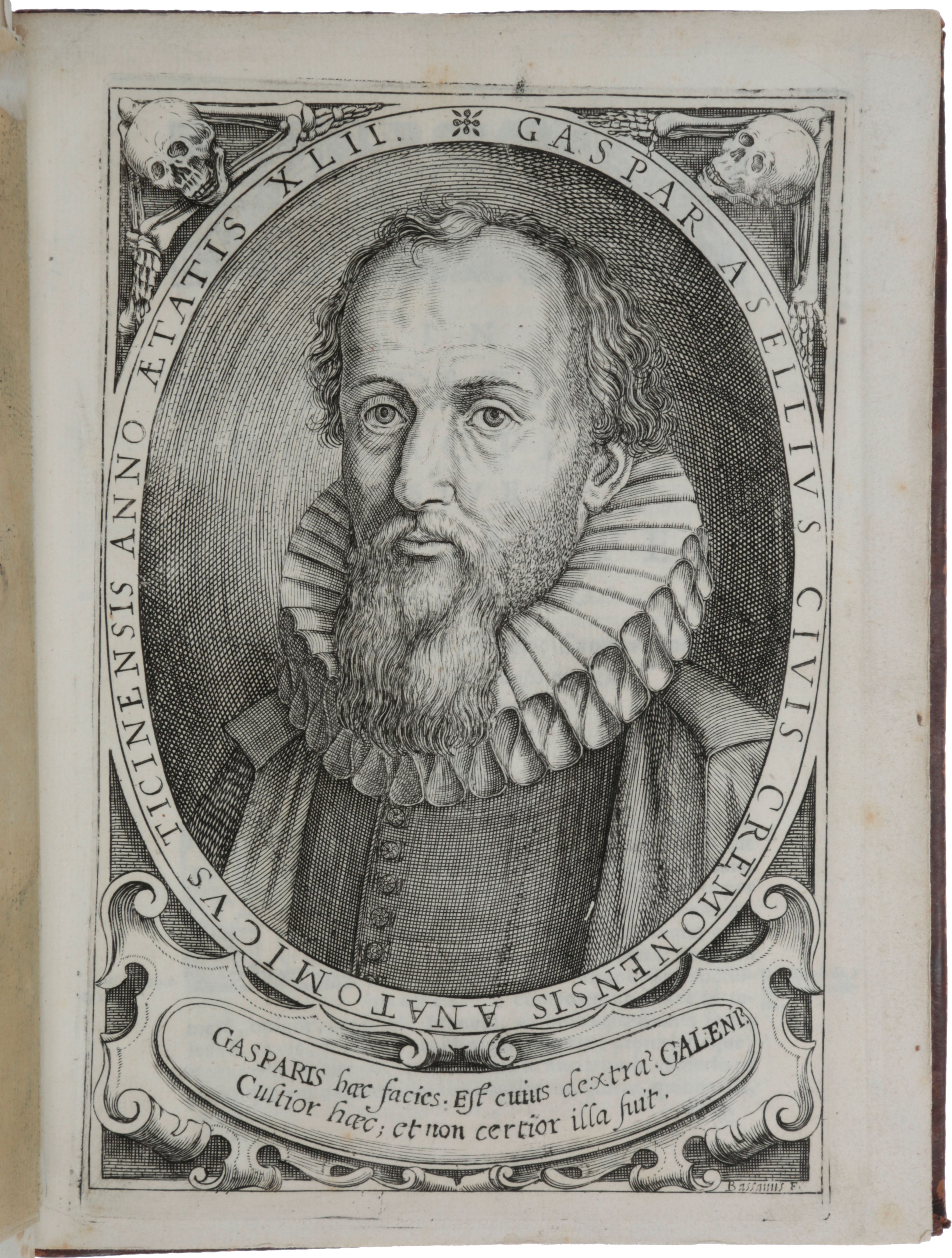
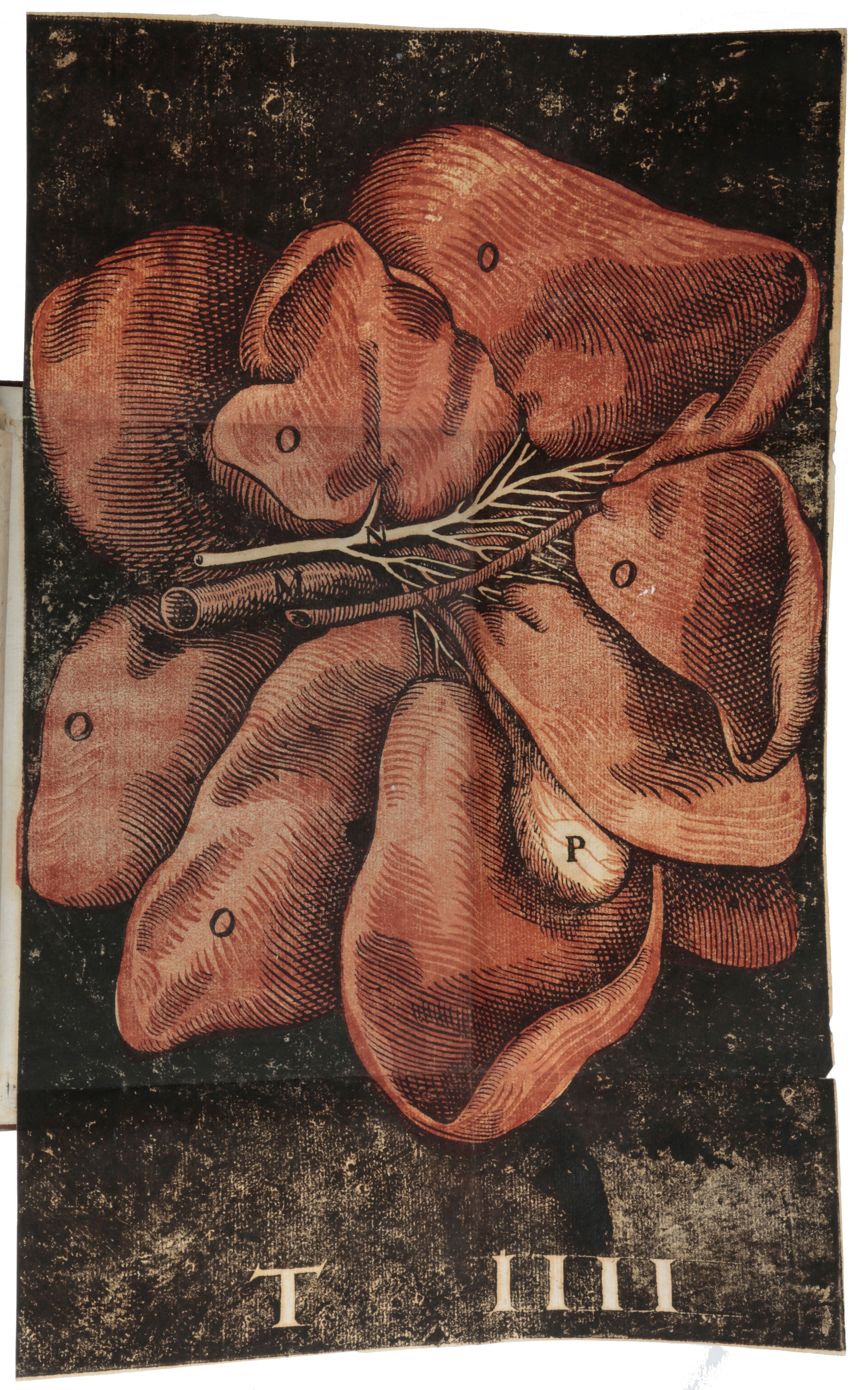
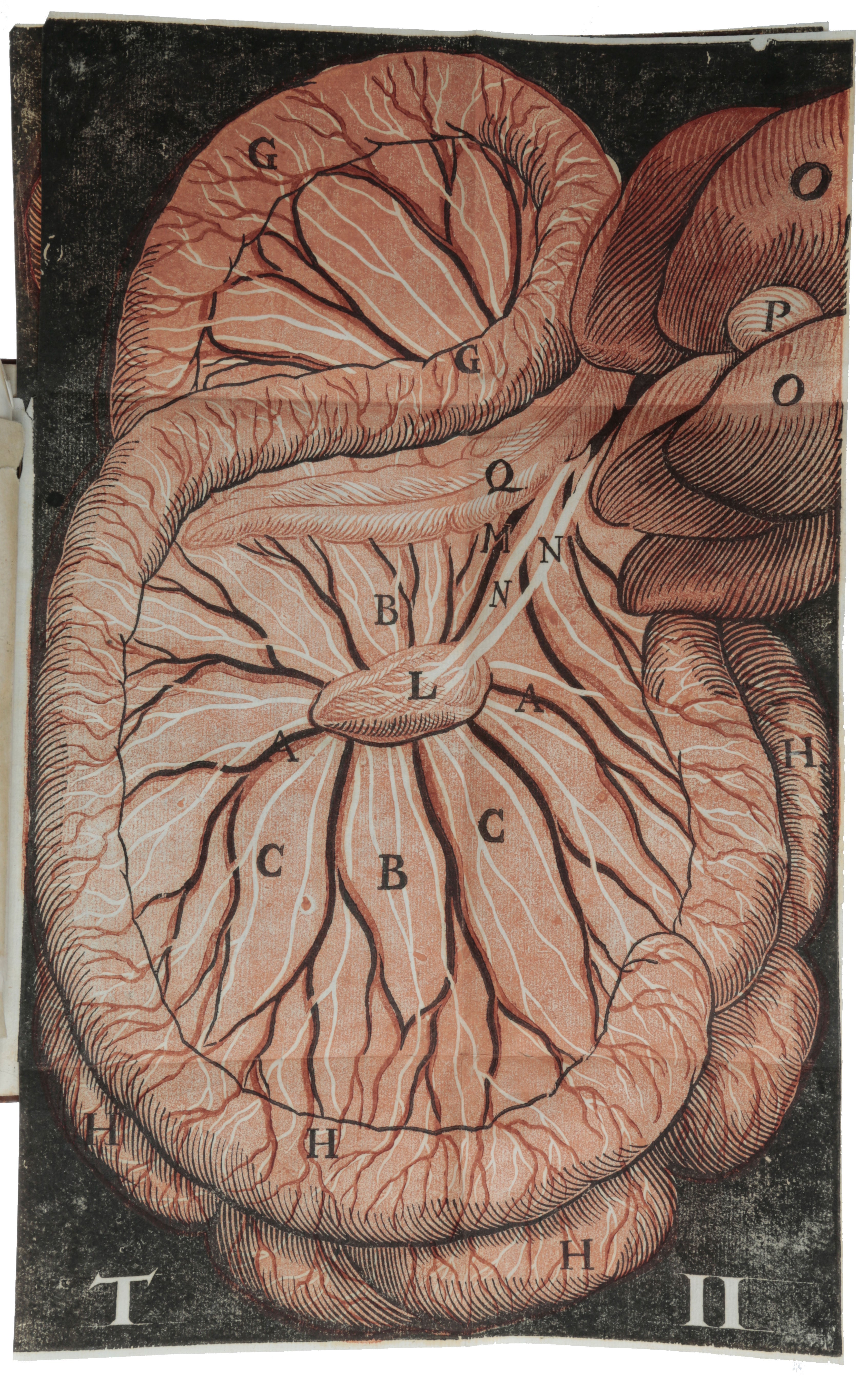
Milan: Giovanni Battista Bidelli, 1627. First edition, a fine copy, of the first book with anatomical illustrations printed in colour. “In 1622, during a vivisection performed upon a dog that had just fed, Aselli (1581-1625) rediscovered the chylous or lacteal vessels, which Galen and Eristratus reported to have been documented by Hippocrates and Aristotle, but which had been overlooked by the anatomists of the sixteenth century. Aselli undertook a systematic study of the vessels in different species of animals, and established the cause and effect relationship of their turgidity with the intake of nourishment: Although he thus recognized their nature and function, he failed to trace them to the thoracic duct, instead mistakenly construing a connection with the liver, still considered the center of the venous system in the decade before publication of Harvey’s De motu cordis. Aselli’s error was rectified in the 1650s, when three scientists working independently, Jean Pecquet, Thomas Bartholin and Olof Rudbeck, nearly simultaneously discovered the thoracic duct. Harvey himself apparently did not know of Aselli’s work” (Norman catalogue). After his death, Aselli’s report of his findings – the only one of his studies to appear in print – was published in Milan at the press of Giambattista Bidelli through the efforts of Nicolas Fabry de Peiresc by his friends Senator Settala and Alessandro Tadino. The work contains a beautiful engraved title page and a portrait of Aselli by the Milanese painter and engraver Cesare Bassano (1584-1648), and four folding chiaroscuro woodcurs printed in black, red, and two shades of brown. In its use of colour printing to more accurately distinguish the different types of vessels depicted, De lactibus was the “first publication to use colored illustrations in the interest of scientific accuracy” (Grolier Medicine). The striking woodcuts, which appear in this edition only, have been attributed either to Cesare Bassano or to his associate Domenico Falcini (c.1575-1632). Subsequent editions were illustrated with black and white reduced and engraved copies of the original woodcuts” (Norman catalogue). ABPC/RBH list six copies. “A descendant of an ancient patrician family, Aselli revealed a marked propensity for the natural sciences early in his schooling. He studied medicine at the University of Pavia, where he soon distinguished himself among his fellow students. His teacher of anatomy was Giambattista Carcano-Leone, a pupil of Fallopio and author of De cordis vasorum in foetu unione (1574). “Later Aselli moved to Milan, where he gained recognition in his profession. Since his scientific preparation was essentially in anatomy, he distinguished himself in his practice of surgery. He was appointed first surgeon of the Spanish army in Italy from 1612 to 1620. In Milan he had the opportunity to continue his anatomical researches, which won him honorary citizenship of that city and an outstanding position in the history of anatomy. He died at the age of forty-four from an acute and malignant fever. “Aselli’s scientific activity occurred during the first decades of the seventeenth century, in an atmosphere that was particularly sympathetic to anatomical studies, especially in northern Italy. At the end of the sixteenth century the study of descriptive human anatomy had made considerable progress. The early decades of the seventeenth century felt the effects of the baroque attitude toward science and the influences of the new mechanical concepts of Galileo. Anatomy, which had been essentially static in the sixteenth century, now assumed a dynamic character. It was enlivened by a new consideration of physiology. “Aselli discovered the chylous vessels, although it would perhaps be more correct to refer to his work as a rediscovery rather than a discovery. According to information that has come down to us from Galen, Herophilus, and Erasistratus, both Hippocrates and Aristotle had with considerable clarity already pointed to the existence of the so-called absorbent vessels. Nevertheless, not even Eustachi and Fallopio, who in the sixteenth century had noted and described the thoracic duct in the horse and the deep lymphatics in the liver, respectively, had succeeded in clarifying the functional significance of these vessels. “On 23 July 1622, during a vivisection performed on a dog that had recently been fed, Aselli, in the course of removing the intestinal tangle to reveal the abdominal fasciae of the diaphragm, noticed numerous white filaments ramified throughout the entire mesentery and along the peritoneal surface of the intestine. The most obvious interpretation was that these filaments were nerves. The incision of one of the larger of these “nerves” released a whitish humor similar to milk. Therefore, Aselli interpreted these formations as a multitude of small vessels, proposing to call them “aut lacteal, sive albas venas.” The vivisection had been performed at the request of four of his friends, among whom were Senator Settala and Quirino Cnogler, to demonstrate the recurrent nerves and the movements of the diaphragm. “Immediately following his discovery, Aselli began a systematic study of the significance of these vascular structures. He recognized the chronological relationship that existed between their turgidity and the animal’s last meal. His experimental findings enabled Aselli to observe the chylous vessels in different species of animals. The results of these investigations were collected in De lactibus sive Lacteis venis quarto vasorum rnesaraicorurn genere novo invento Gasparis Asellii Cremonensis anatomici Ticinensis dissertatio (1627), which is divided into thirty-five chapters that are followed by an index and preceded by four charts with accompanying commentaries and a portrait of the author. Besides their intrinsic scientific value, the importance of the charts lies in the new technique used in composing them: they were the first anatomical illustrations to appear in color. Aselli used color because he felt ihat several tints were needed in order to distinguish the various types of vessels more clearly. “Aselli traced the course of the chylous vessels to the mesenteric glands and probably confused them with the lymphatics of the liver; therefore he did not follow their course to the thoracic duct. (His discovery occurred six years before the publication of Harvey’s De motu cordis, and so the Galenic concept of the liver as the center of the venous system still appeared valid.) Harvey himself believed that the absorption of the chyle took place through the mesenteric veins and that the liver generated the blood. With the discovery of the thoracic duct in the dog by Jean Pecquet in 1651, the old Galenic error, according to which the vessels of the intestine carried the chyle to the liver, was corrected. In addition to noting and describing the valvular apparatus of the chylous vessels, Aselli attempted to interpret their functional significance in health and in disease” (DSB). “It is firstly worth noting that Aselli’s work follows Fabricius’ format for anatomical reports, that consists of a three-section structure: historia, actio, utilitas. Anatomists should firstly provide a full description of the concerned body part, without any tentative reference to causes. This means that the first section, called ‘historia,’ focuses only on the structure of the part. Secondly, anatomists should proceed with ‘actio,’ that is the description of the action of the part. Finally, the last section, ‘utilitas,’ should offer the account of the final causes, that is, the reasons why that part exists. “In the section devoted to historia, after describing the main mesenteric vessels, Aselli provides a detailed report of his accidental discovery (‘De Quarto, & novo Vasorum Mesairacorum genere’). On July 23, 1622, Aselli dissected a post-prandial dog, in order to see the recurrent nerves and the movements of the diaphragm. However, the opening of the abdomen and the displacement of the intestines suddenly revealed something completely new, that is ‘a great number of slender cords, so to speak, extremely thin and white, scattered all over the whole mesentery and intestine, from almost innumerable starting points’. Aselli believed those ‘cords’ to be nerves, but then he realized that this hypothesis was untenable. Influenced by the scholarly disputes on meseraic veins, he decided to lance one of those whitish filaments so as to look at it inside. He noticed a milky liquid filling in, a feature that suggested to him the name of ‘venae albae et lactae.’ Then, in support of his discovery, he dissected another dog, but without finding the new vessels. But, since this second dog was unfed, he inferred that there was a correspondence between the food intake and the chance to detect the chyliferous vessels. Thus, he tried again with a third dog, but after six hours from the animal's last meal. Indeed, the vessels reappeared. “Nevertheless, despite the novelty of his discovery, Aselli continues to support Galen’s physiology: as a consequence, chyliferous vessels are considered to lead to the liver, which inevitably preserves its privileged position as the organ of sanguinification … In particular, by describing the progressus of chyliferous vessels, Aselli confused a large mesenteric lymphatic gland with a pancreas of sorts (now known as Aselli’s pancreas or Aselli’s gland), from which larger vessels were supposed to reach the caval vein, the portal vein, or directly the liver … Evidently, the idea of chyle flow which follows from this explanation of the action and the use of chyliferous vessels prevented Aselli from correctly finding the thoracic duct, which is the necessary step to make Galenic view's refutation possible. In fact, it is necessary to show that the chyle cannot reach the liver. “Numerous historians of medicine agree to highlight the contraposition in Aselli’s work between personal experiences by means of direct observations and dissections and the provided theoretical explanations. In this regard, for example, Portal said that ‘L'Auteur se perd dans une théorie qui n'est appuyée sur aucun principe de physique’: Aselli would not have supported Galen's theory, if he had been aware of the existence of the thoracic duct, which was already observed by Eustachi in horses. ‘En adaptant la découverte de ce grand homme à la sienne,’ he wrote, ‘Asellius eût conduit le chyle dans la veine souclaviere gauche, & non dans le foie’. Similarly, in the 19th century, Charles Daremberg said: ‘Encore une fois, voilà ce que produit la mauvaise physiologie ou la physiologie a priori, la physiologie non expérimentale. Le hasard fait trouver les chylifères, la théorie galénique fait qu'on les voit se terminer au foie”” (Tonetti, pp. 71-72). Choulant-Frank, pp. 240-241; Garrison-Morton 1094; Grolier Medicine 26; Heirs of Hippocrates 453; NLM/Krivatsy 446; Osier 1846; Wailer 502; Wellcome 6837; Norman 76. Tonetti, ‘The discovery of lymphatic system as a turning point
in medical knowledge: Aselli, Pecquet and the end of hepatocentrism,’ Journal of Theoretical and Applied Vascular Research 2 (2017), pp. 67-76.
4to (224 x 170 mm), pp. [xxiv], 79, [1], including engraved title, engraved author portrait, both by Cesare Bassano (both conjugate with text leaves), 4 large folding chiaroscuro woodcut plates printed in black, red and brown, woodcut initials and head-piece. Contemporary speckled calf, double gilt fillets on boards, spine ruled in gilt with gilt lettering-piece (a little rubbed, joints starting).
Item #5795
Price: $115,000.00
